Beyond diagnostic accuracy, artificial intelligence, integrated with modern healthcare software, allows medical staff to focus on complex cases, improving the state of overall patient care.
Keep reading the article to learn more about how medical imaging improves with the help of AI, its applications, and use cases.
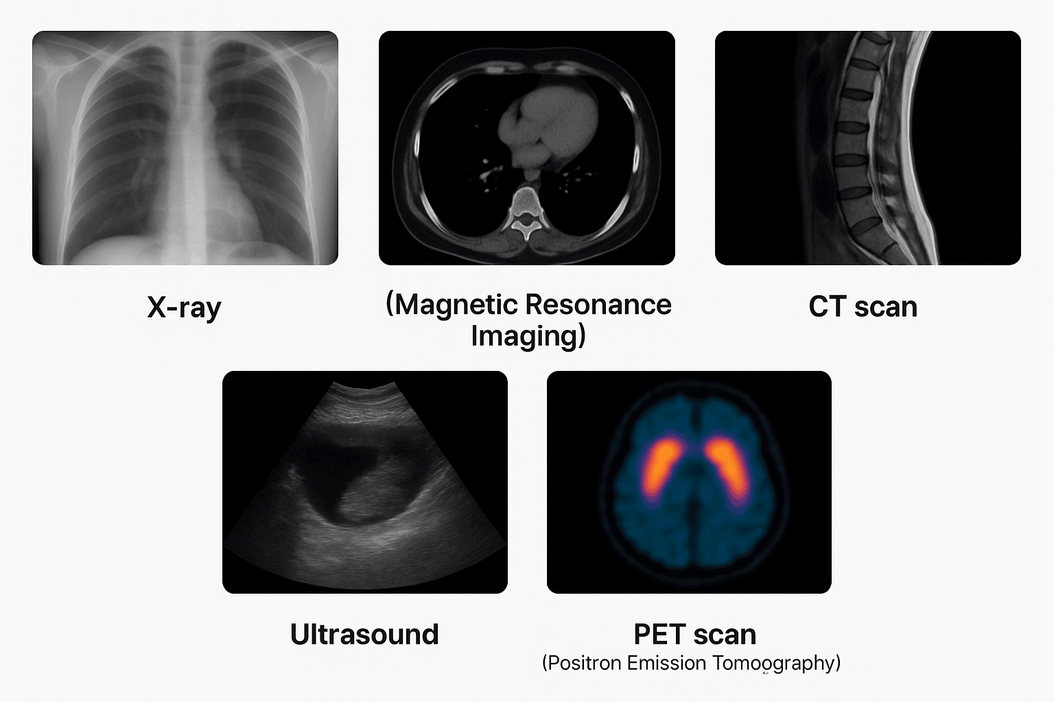
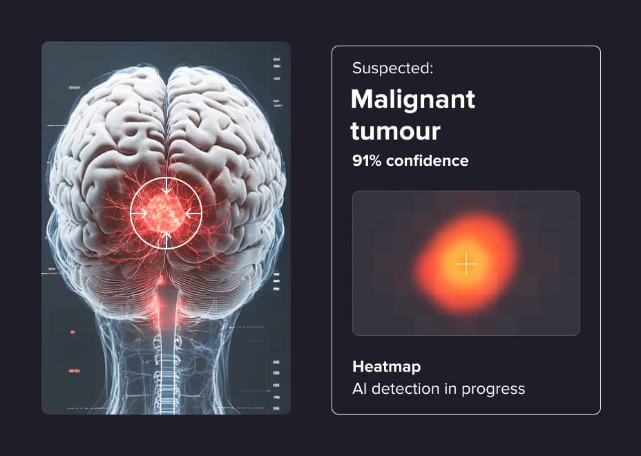
AI can detect diseases in medical images very accurately. It can identify a tumour in the lungs or changes in the eye at early stages. This means diseases can be detected earlier, allowing treatment to start sooner. For example, AI-based convolutional neural networks (CNNs) not only matched the diagnostic sensitivity of experienced radiologists but also identified 8.4% of lung nodules that would have otherwise been missed in patients with complex lung conditions.
AI processes images much faster than humans. As a result, doctors receive results more quickly. This is especially important in emergencies, where every second is counted. MedSAM2 (Medical Segment Anything Model 2) is a foundation model for 3D medical image and video segmentation, trained on over 455,000 3D image-mask pairs and 76,000 video frames. With a human-in-the-loop pipeline, it reduces manual annotation effort by over 85% and accelerates workflows.
AI can accurately measure how much a tumour has grown or shrunk and how the patient's condition changes over time. The doctor sees not just an image but specific numbers, which helps to better assess whether the treatment is working and make changes over time.
In rural areas where access to medical specialists is limited, AI assists doctors by analysing medical images and highlighting potential issues. This support makes high-quality healthcare more accessible and consistent for underserved communities.
Role of machine learning in medical imaging
Machine learning (ML) is the technology that primarily teaches the empty model to make decisions or predictions based on data that has such knowledge (historical data, previous decisions, labelled content, etc.). For example, you can predict some value in the future based on the series of historical values, classify images based on image labels set by humans in advance, and generate text based on huge corpora (books, forums, articles) seen by the model.
Traditional machine learning relies more on clearly defined features and rules created by data scientists, while deep learning uses complex neural networks to automatically learn from large amounts of imaging data.
Machine learning techniques are often easier to understand and interpret, making it simpler to explain why a certain diagnosis or prediction was made. This helps doctors trust the results and make better decisions. Machine learning also works well with smaller datasets and can be combined with other patient information, offering practical tools to improve clinical care alongside deep learning methods.
Deep learning techniques in medical imaging
Deep learning (DL) is the technology that focuses on the Deep Neural Nets (MLPs, CNNs, RNNs, Seq2Seq, GANs, Transformers, Diffusion models, KANs, etc.) usage as the primary models, but for the same tasks from the forecasting and ending with the text generation.
Deep learning has made major progress in recent years due to the ability to learn complex patterns and features from raw data. In healthcare, DL systems can also check for mistakes or unusual patterns in medical data. They can even verify whether medical images are real or have been tampered with—an important feature in the digital transformation era. DL can also predict how a disease might progress or how a patient may respond to treatment.
Deep learning uses different neural networks for many medical imaging tasks:
- Convolutional Neural Networks (CNNs) learn features from images using layers that detect patterns, reduce data size, and classify content, making them highly effective for tasks like image recognition and medical image analysis.
- Recurrent Neural Networks (RNNs) are deep learning models designed to process sequential data. They use internal memory to retain information from previous steps, making them ideal for evaluating a series of images from slices (MRI) or from different timeframes.
- Generative Adversarial Networks (GANs) generate realistic data by training two networks, a generator and a discriminator in competition. The generator creates fake data; the discriminator tries to detect if it's real or fake. In imaging tasks, GANs are used for synthetic data generation and image enhancement.

View all case study
Current challenges in medical imaging
1. Low-quality medical images
Medical images can sometimes suffer from reduced quality due to various factors: the skill and experience of the operator, patient movement or positioning, and specific imaging conditions.
AI solution
AI learns by looking at many pairs of blurry and clear versions of the same picture. By studying these pairs, it figures out how to turn a blurry image into a clearer one. After learning this, the AI can make low-quality images sharper, improve their detail, and keep important things like tumours or small changes in tissue visible.
2. Radiologist shortage and diagnostic delays
Modern medicine uses more imaging tests than ever before. The delay is typically caused by the chain of doctors' reviews (diagnose/radiology technologist is different from pure radiologist and therapist). So, according to medical protocols, the diagnosis is provided by different doctors with different qualifications. As a result, patients may wait a long time for results, even when every minute is critical.
AI solution
AI tools can quickly scan medical images and spot signs of serious problems like bleeding or tumours. When they find something dangerous, they can automatically send it to a doctor for an urgent review.
This helps doctors deal with the most critical cases first and shortens the time patients wait for answers in emergencies.
3. Variability in interpretation
The interpretation of medical images depends on the experience and knowledge of radiologists. Even experienced doctors can evaluate the same images differently; some may spot small lung nodules that others might overlook.
AI solution
Artificial intelligence analyses medical images objectively in a standardised way. AI algorithms identify suspicious areas and act as a “second reader”. This helps make image interpretation more consistent and:
- Identify abnormalities that a radiologist might have missed
- Provide exact measurements to support and enhance visual evaluations
- Ensure consistent and accurate results, regardless of fatigue or workload
4. Data fragmentation and privacy concerns
Medical imaging data is scattered across many hospitals and stored in different formats. Strict privacy regulations (like HIPAA and GDPR) restrict the sharing of patient data. This fragmentation makes it difficult to develop accurate AI models, which need large and diverse datasets. One of the possible solutions to this problem is data anonymization.
Generalised approach
AI processes data at each hospital without sharing private patient information. Only the AI’s updates are shared, keeping data safe while learning from many sources. Supporting this secure approach, ELEKS has earned HITRUST e1 Certification, proving its ability to protect healthcare data and deliver compliant solutions like Compliance Automation Platform (eCAP), trusted by U.S. healthcare providers.
5. Challenges in AI implementation
AI models may produce false positives and hallucinations or exhibit bias, which can compromise the reliability of their outputs. As a result, their performance must be carefully verified and validated with subject matter experts (SMEs) to interpret results and check if the model's decisions align with clinical or domain-specific standards.
As AI becomes more integrated into diagnostic imaging, it helps address key challenges in the field. However, this sensitivity also brings a challenge—the technology may detect subtle changes that doctors don’t think are serious. For instance, AI systems might flag tiny lesions in a scan that a radiologist will typically ignore. These findings, sometimes referred to as "incidentalomas," can lead to overdiagnosis and unnecessary follow-up tests, biopsies, or treatments, increasing patient anxiety and burdening healthcare systems.
Conclusion
Despite some challenges, AI in healthcare shows promising results in medical imaging and a strong potential to transform the field. It is important to have teamwork between doctors, AI developers, and regulators to tackle ethical and practical issues while making the most of what AI can offer.
AI also helps improve the speed and accuracy of diagnoses, treatments, personalisation, and how care is delivered overall. It also makes results clearer and easier to understand and supports better communication among healthcare teams.


Related Insights


The breadth of knowledge and understanding that ELEKS has within its walls allows us to leverage that expertise to make superior deliverables for our customers. When you work with ELEKS, you are working with the top 1% of the aptitude and engineering excellence of the whole country.

Right from the start, we really liked ELEKS’ commitment and engagement. They came to us with their best people to try to understand our context, our business idea, and developed the first prototype with us. They were very professional and very customer oriented. I think, without ELEKS it probably would not have been possible to have such a successful product in such a short period of time.

ELEKS has been involved in the development of a number of our consumer-facing websites and mobile applications that allow our customers to easily track their shipments, get the information they need as well as stay in touch with us. We’ve appreciated the level of ELEKS’ expertise, responsiveness and attention to details.